
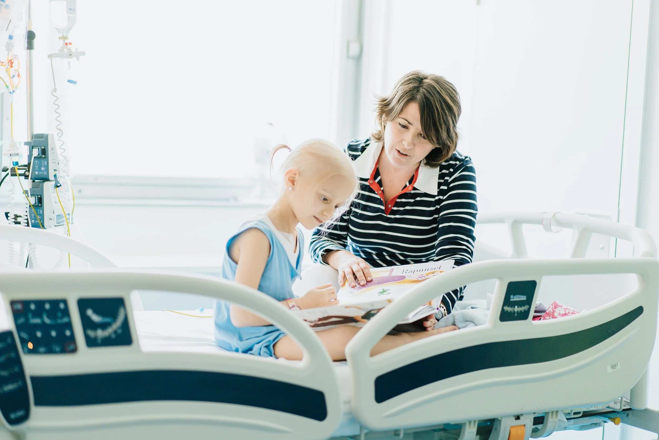
PAEDIATRIC PROJECTS
Childhood cancer is a rare disease, yet it continues to receive disproportionately low levels ofresearch funding.
At CRIS Cancer Foundation, every child counts. That is why we are committed to driving pioneering research programmes that aim to change the outlook for children diagnosed with the most challenging cancers, supporting innovative projects and collaborations with leading hospitals, universities, and research centres around the world.
We accelerate the development of new treatments through translational research, precision medicine, immunotherapy, and next-generation therapies, giving every child with cancer the best possible chance of a healthy future
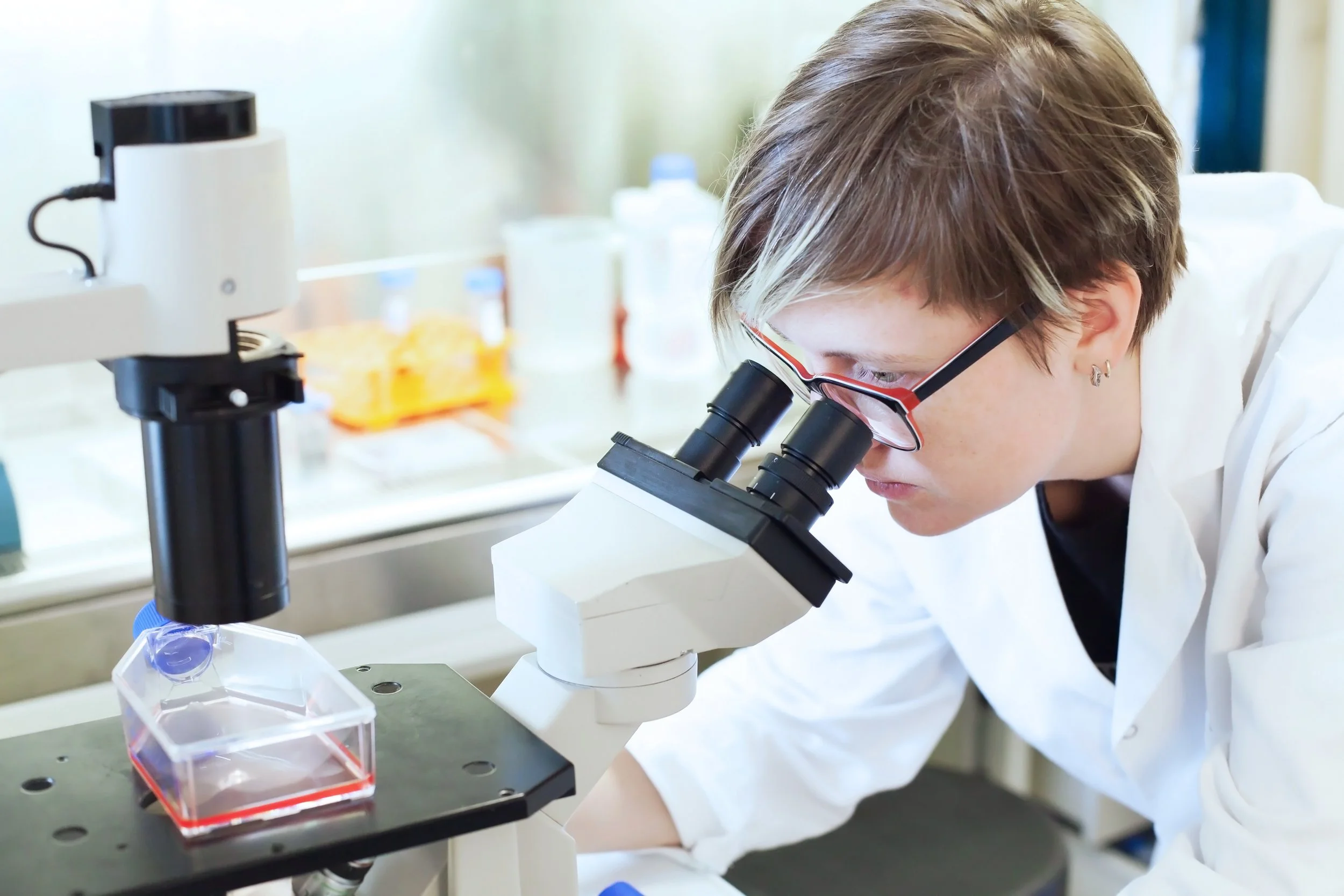
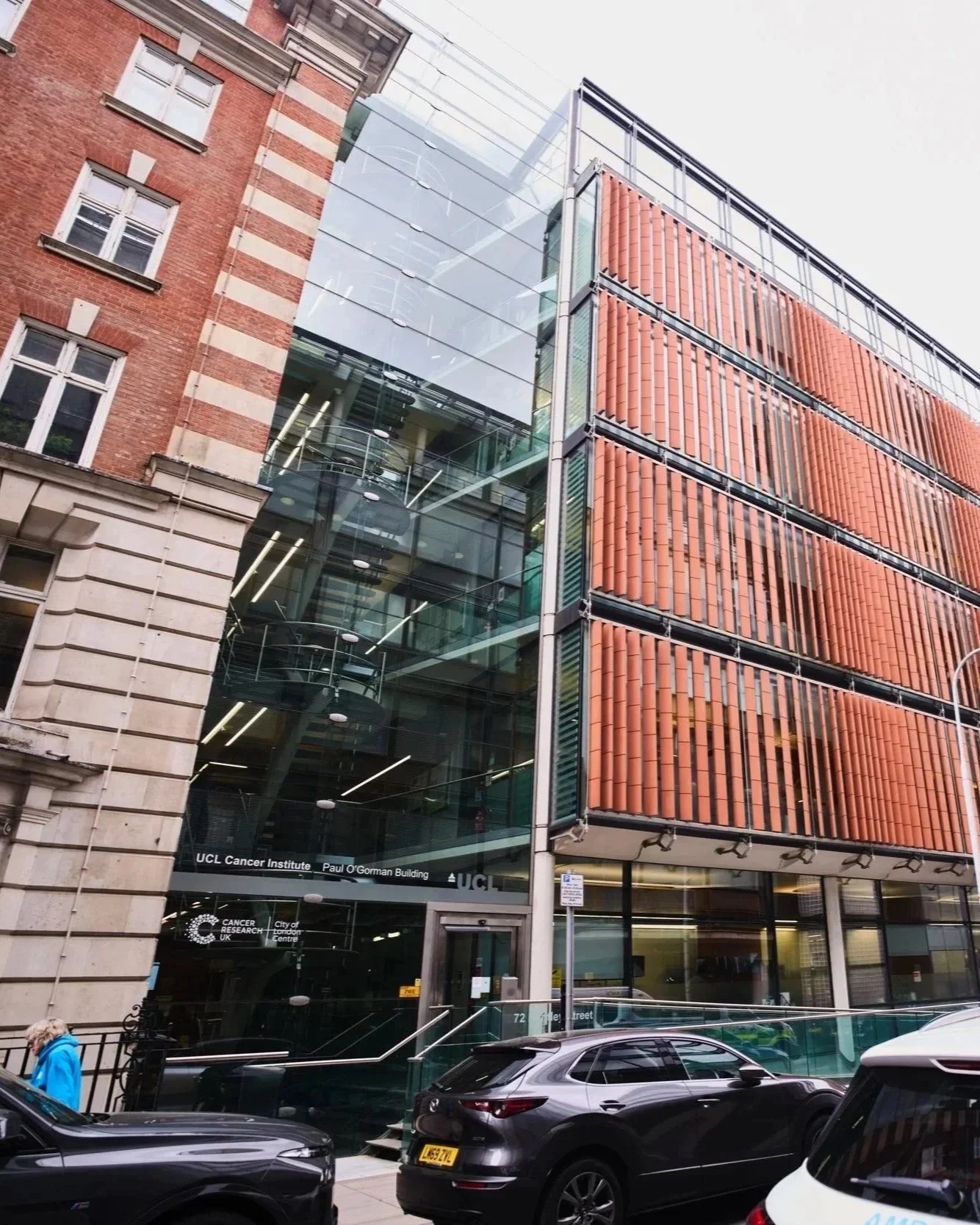
The Institute of Cancer Research (ICR)
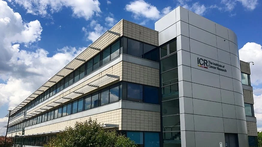
The Institute of Cancer Research (ICR), London, is a leading centre focused on understanding cancer and developing new treatments. Alongside its work in adult cancers, it is also addressing paediatric cancers, which have distinct challenges and treatment needs.
Through CRIS Cancer Foundation’s support, research at the ICR is helping improve options for children who do not respond to standard treatments, with a focus on developing more effective and less toxic therapies, including targeted treatments and immunotherapy.
-
Principal Investigator: Prof Chris Jones and Dr Matthew Clarke
Centre: ICR, London
Introduction:
Pediatric brain tumors remain one of the greatest challenges in modern medicine. This is due not only to their biological heterogeneity but also to the poor prognosis that many of these tumors carry. Their diagnosis is often a devastating event for families, as effective treatments are still lacking for some tumor types.
Although globally around 70% of children diagnosed with brain cancer survive more than five years (according to the CONCORD-3 study), for some tumor types this percentage drops to only about 20% (according to the American Cancer Society). For certain tumors there are still very few effective treatments available, and the average survival after diagnosis may sometimesbe no more than a year.
In addition, the treatments that are currently available often need to be very aggressive, and they can leave patients with severe long-term side effects.
For this reason, it is essential to improve diagnostic methods and to identify the specific characteristics of each patient’s tumor so that more specialized and personalized therapies can be applied. Pediatric brain tumors are very different from those that occur in adults and have distinct biological characteristics that mean they cannot be treated in the same way.
If we are able to identify specific features or vulnerabilities in the tumors of each child, doctors can select the most appropriate therapy, greatly increasing the chances of success while avoiding the side effects of treatments that may not even be effective.
The Project
Professor Chris Jones, from the Institute of Cancer Research in London (UK), works to identify a wide range of molecular alterations—genetic changes as well as other cellular modifications—in pediatric brain tumors. The goal is to better distinguish between tumor types, particularly some of the most aggressive forms, such as Pediatric High-Grade Glioma, Diffuse IntrinsicPontine Glioma (DIPG—the tumor type studied in the CRIS Brain Tumor project in France), and Diffuse Midline Glioma (DMG—the focus of a clinical trial supported by CRIS at Great Ormond Street Hospital).
This research helps scientists better understand the biological processes that cause a cell to lose control and develop into a tumor. Ultimately, this knowledge can improve patient prognosis and help clinicians select the most appropriate treatment.
An important aspect of this work is improving the precision of classification methods for pediatric brain tumors. If patients with tumors that share similar appearances and genetic alterations can be grouped together, each group can potentially be treated differently. The more precisely these disease groups are defined, the more targeted and effective treatments can become.
Professor Jones’s group is a European leader in this type of research, and most of their studies involve collaborations across many different countries.
Moreover, a deeper molecular understanding of these tumors may also reveal specific weaknesses that can be targeted therapeutically.
A clear example of this approach is a study published in 2020 in the prestigious journal Nature Communications, where the team observed that patients under the age of four with Diffuse Intrinsic Pontine Glioma (DIPG) often present mutations in a protein called ALK2. Drugs that target this protein already exist, and treatment with these drugs was able to reduce tumor size in some patients.
Similarly, in a study on Diffuse Midline Glioma published in the international journal Cancer Discovery, the team identified a drug combination (vandetanib and everolimus) that showed excellent results in laboratory models. This combination was later used on a compassionate-use basis in four patients from different parts of Europe, and in one of them it prolonged life for 20 months with a good quality of life.
Current Research Lines
The group’s current research focuses on several main areas:
Study of genetic and epigenetic alterations in pediatric brain tumors
Many advances in the diagnosis and treatment of these tumors have come from genetic analysis—studying mutations in the DNA of tumor cells. However, not all relevant changes are strictly genetic. Some involve alterations in how cells read and interpret the information contained in DNA. This is known as epigenetics.
Studying these epigenetic changes alongside genetic information allows researchers to deepen their understanding of these tumors, refine diagnostic classifications, and potentially design new therapeutic strategies.
Analysis of long-term survivors
Although children with high-grade gliomas or DIPG typically have very short survival times, a small number of patients survive much longer. Dr. Jones’s team is studying the characteristics of these patients to understand why they survive while others do not.
The aim is to use this knowledge to develop new, better-targeted therapeutic strategies that could improve survival for children with the poorest prognoses.
Development of robust laboratory models
Laboratory models that accurately reproduce tumor behavior are essential for studying these cancers and developing new treatments. To address this need, Dr. Jones’s team is building an extensive collection of three-dimensional cell cultures derived from tumors from a large number of patients.
Study of the spatial characteristics of pediatric brain tumors
For many years, tumor research focused mainly on measuring how much DNA, RNA, or protein was present in a tumor sample. However, recent studies have shown that where these molecules are located within the tumor is just as important as how much is present.
To explore this dimension, the team is using highly advanced tools to understand the characteristics of tumor cells in different regions of the tumor, as well as their interactions with the surrounding
Recent Advances
Dr. Chris Jones’s team continues to be an international reference in the study of the most aggressive pediatric brain tumors. One of their earlier studies, focused on the molecular characterization of these tumors, has become one of the most widely cited publications in the field, with more than 900 citations in scientific literature—an indication of its enormous impact and usefulness for the medical and scientific communities.
With support from CRIS, the team has achieved several significant advances in the past year.
Improved diagnostic classification of pediatric brain tumors
One of the major challenges in pediatric brain tumors is that they are not a single disease but rather many different diseases that until recently were treated in almost identical ways. This partly explains why treatments sometimes fail: a therapy that works for one tumor type may have no effect—or even be harmful—for another.
To move toward more precise and effective treatments, it is essential to determine exactly which tumor type each patient has.
Dr. Jones’s team has analyzed more than 2,300 samples of pediatric gliomas using cutting-edge technologies such as genetic sequencing, epigenetic profiling, and gene expression analysis. This work has allowed them to refine the classification of these tumors and identify new subtypes with distinct characteristics.
This improved classification makes it possible to:
Achieve more precise diagnoses from the outset
Better predict each patient’s prognosis
Select more appropriate therapies for each tumor subtype
Design more effective clinical trials focused on well-defined patient populations
This advance has direct clinical implications: it opens up new opportunities to improve treatment for children facing these aggressive diseases, where therapeutic options are often limited.
Understanding the tumor in its spatial context
For many years, tumor research focused on identifying which molecules are present in tumors—DNA, RNA, and proteins—but not on where these molecules are located within the tumor tissue.
Today we know that the spatial organization of cells within tumors plays a crucial role in determining how tumors behave, how tumor cells interact with one another, and why they respond—or fail to respond—to treatment.
Chris Jones’s team has applied cutting-edge spatial analysis techniques to study the internal organization of these tumors. In certain subtypes, such as gliomas with mutations in the H3G34 gene, they discovered that tumor cells are not randomly distributed. Instead, they form complex structures that resemble stages of normal brain development.
These structures consist of “nests” of tumor stem cells surrounded by more immature cells, suggesting that the tumor recreates an environment that promotes its own growth.
These findings help researchers:
Understand how the tumor originates and sustains itself
Identify key regions within the tumor that may be more vulnerable to specific treatments
Develop strategies to disrupt communication between tumor cells, one of the mechanisms underlying their resistance and aggressiveness
In summary, this spatial approach is opening new therapeutic opportunities for some of the most complex pediatric brain tumors.
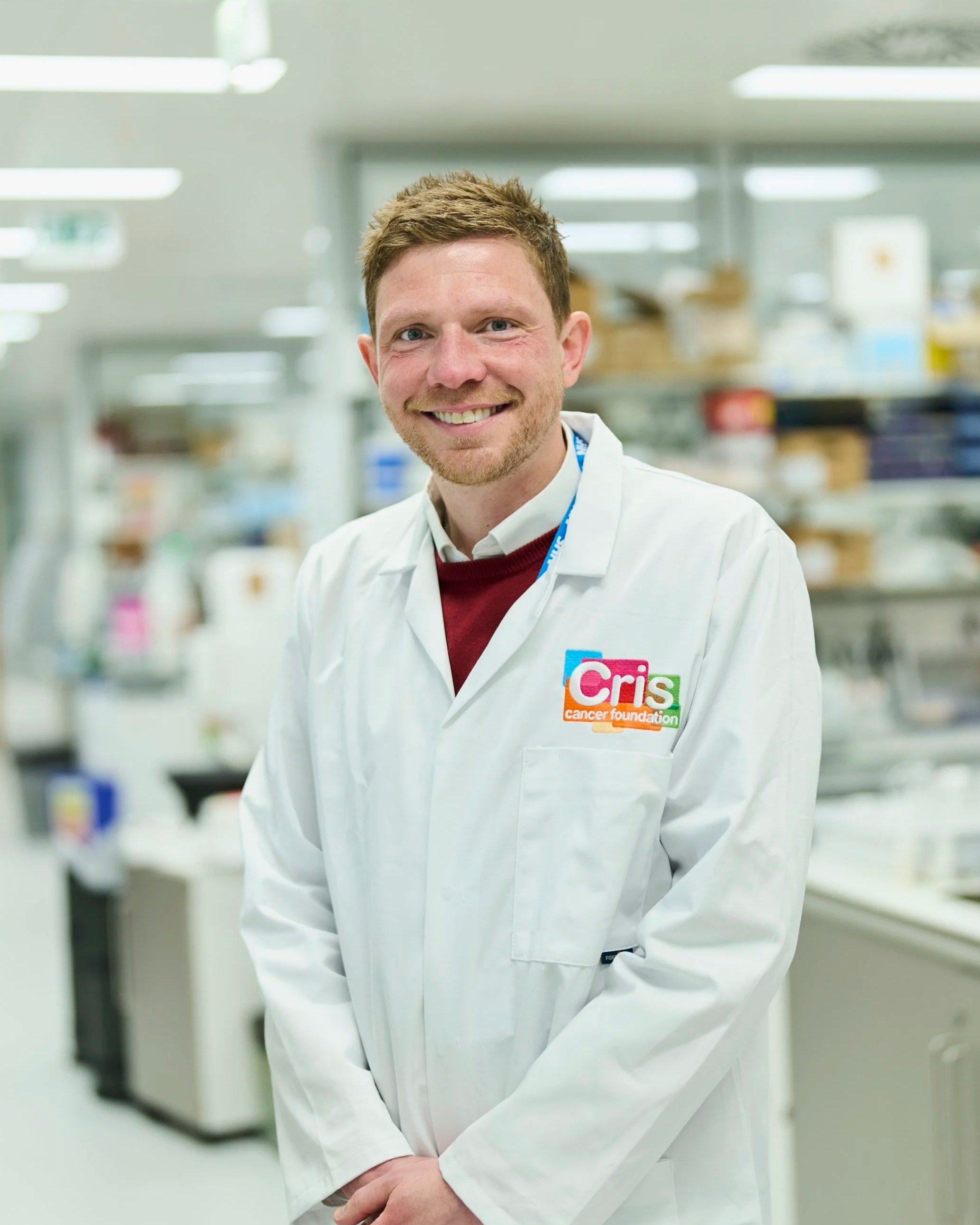
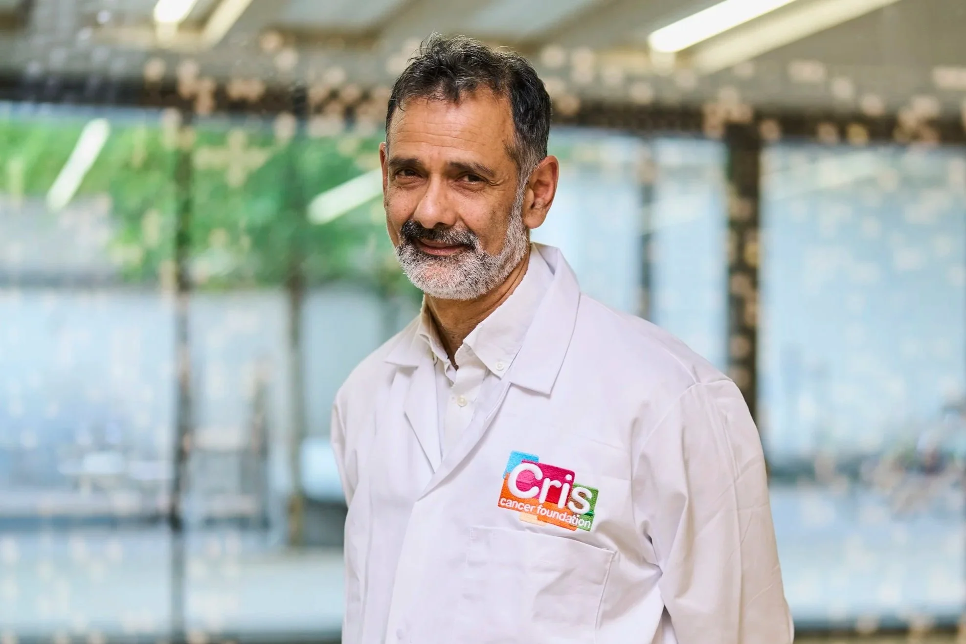
Prof Chris Jones and Dr Matthew Clarke
-
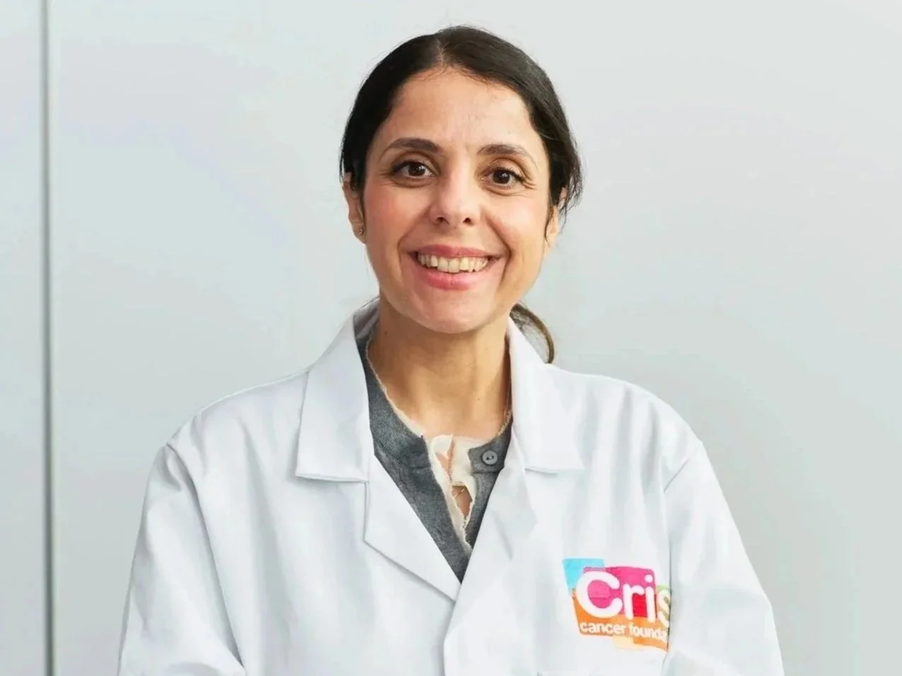
Researchers: Prof Janet Shipley
Centre: ICR, London
Introduction
Paediatric rhabdomyosarcomas are a type of tumour that develop in children’s muscle tissue during growth. They account for around 8% of all childhood cancers, making them the most common soft tissue sarcomas in children and a significant cause of cancer-related death in childhood. This is due to their highly aggressive nature: when they do not respond to treatment or spread throughout the body, survival rates fall below 30%.
In these situations, treatment options are unfortunately very limited, and survival has not improved significantly in recent years. Moreover, existing treatments can leave serious long-term side effects. Even approaches such as immunotherapy, which have transformed outcomes in other childhood cancers, have shown limited success in rhabdomyosarcoma. This is because these tumours can evade the immune system, effectively becoming “invisible” — they display very few signals that would allow the body’s defences to recognise and attack them.
There is therefore an urgent need for new strategies and treatments to provide better options for the most severe cases of rhabdomyosarcoma.
The Project
Researchers Janet Shipley and Zoe Walters are experts in rhabdomyosarcoma. In this project, they aim to help the immune system recognise and destroy tumour cells associated with this cancer.
To achieve this, they will use epigenetic strategies — approaches that modify how cells read the instructions encoded in their DNA — specifically targeting tumour cells. They will develop advanced laboratory models that replicate the tumour environment and test two treatments that are already known and used in other paediatric cancers: one targeting a protein (EZH2) that helps the tumour remain hidden, and another using retinoic acid, which is employed in other childhood cancers to make tumour cells less aggressive.
These treatments could transform an “invisible” tumour into one that is highly visible to the immune system, enabling it to detect and eliminate cancer cells more effectively, while also enhancing the impact of immunotherapies that boost immune cell activity.
This approach could open the door to new, effective, and safer treatments for children whose options are currently very limited.
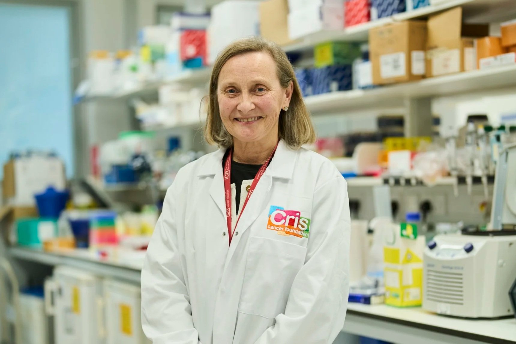
Prof Janet Shipley
-
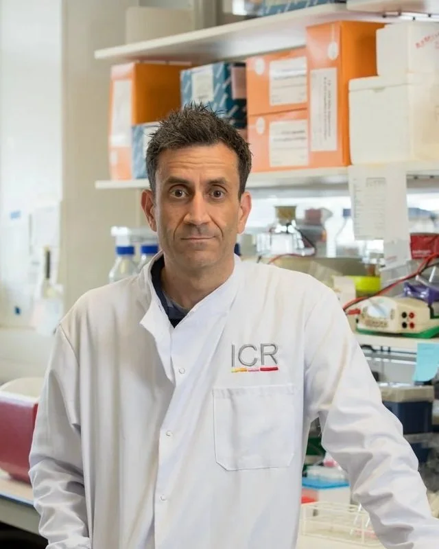
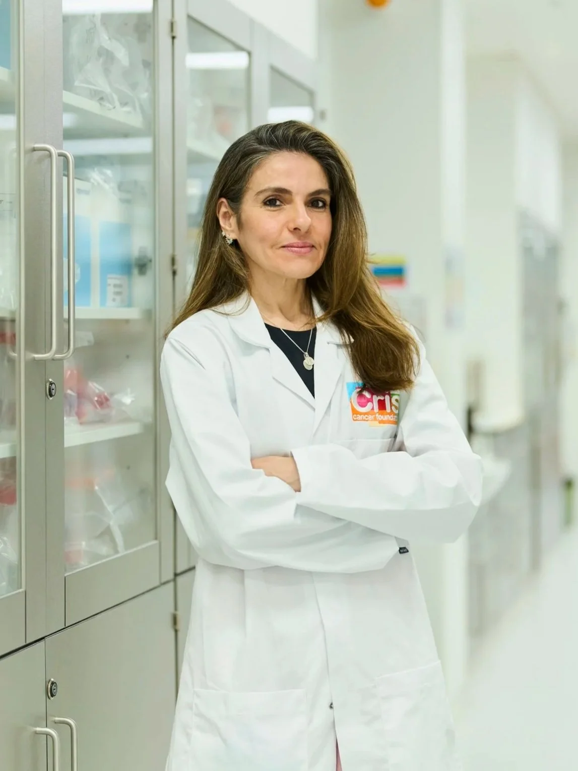
Researchers: Prof Sir Mel Greaves and Dr Marcela Braga
Centre: ICR, London
Introduction
Thanks to ongoing research, major advances have been made in recent years in targeted therapies, immunotherapy, and combination treatments, and cancer survival rates continue to improve. However, when cancer reaches advanced or metastatic stages, it still poses a major challenge and, in many cases, remains very difficult to cure. In these situations, although initial treatments may reduce tumour size or even eliminate it, many patients eventually relapse over time.
The main reason for these relapses is the emergence of resistant tumour cells that escape the effects of therapy, survive, and eventually begin to multiply again. Therefore, one of the key challenges in modern medicine is to understand when and how this resistance develops. If we can uncover these mechanisms, we could potentially prevent resistance, improving the lives of millions of patients worldwide.
It is now known that tumour cells evolve over time and can adapt to hostile conditions, including medical treatments. However, it remains unclear why some cells do so with such efficiency. All cells—both healthy and cancerous—contain a gene that acts as a safeguard when DNA is damaged: TP53, often referred to as the “guardian of the genome.” When TP53 detects problems, it can halt cell division or trigger cell death, making it a powerful defence against cancer.
However, some tumour cells lose TP53. This not only prevents the cell from dying when something goes wrong but, according to a recent hypothesis, may also enable these cells to evolve more rapidly—acting like “evolutionary machines” capable of diversifying and adapting more effectively to treatments. Although this theory is compelling, it has not yet been fully demonstrated in the laboratory.
The Project
Dr Marcela Mansur Braga, part of the team led by the renowned Dr Mel Greaves—one of the world’s leading researchers in leukaemia—has launched an ambitious project to test whether the loss of the TP53 gene not only promotes tumour cell survival but also accelerates their evolution. This rapid evolution could allow tumour cells to generate high levels of diversity, increasing the likelihood that some will become resistant even to future treatments.
To investigate this hypothesis, she will use highly advanced laboratory models based on leukaemia cells that have been genetically engineered to either retain or lack TP53. Using sequencing techniques, cellular analysis, and animal models, she will study how these cells change over time and whether those without TP53 are more likely to develop resistance.
If this theory is confirmed, it could open up a new approach to anticipating resistance, enabling earlier identification of the most dangerous tumours and the development of treatments designed to prevent resistance before it even arises. A truly visionary project that could represent a major step forward in cancer therapy.
Prof Sir Mel Greaves and
Dr Marcela Braga
Oxford University

CRIS Cancer supports leading cancer research at the University of Oxford by advancing innovative approaches that bridge scientific discovery and clinical care. This work focuses on developing new strategies for cancer diagnosis, treatment, and patient outcomes through cutting-edge research and real-world application.
-
Researcher: Dr Rebecca Ling
Institution: University of Oxford, Oxford
Introduction:
Acute myeloid leukemia in children is a rare but highly aggressive disease, and treatment options remain limited. Unlike other types of childhood leukemia, therapeutic advances have been slower, and many patients require very intensive treatments that can leave significant long-term side effects.
In some cases, leukemia begins even before birth, during fetal development. This means that the cells that give rise to the disease are not the same as those found in an older child or an adult, and that the moment at which the genetic alteration associated with this disease appears may be key to understanding why these tumors are so difficult to treat.
Understanding when and how leukemia begins is essential for finding new ways to target it more effectively and safely.
The Project
Paediatric acute myeloid leukaemia is a rare and highly aggressive disease, particularly in infants, with limited treatment options. This project, led by Dr Ling, investigates whether the developmental stage at which the genetic alteration arises (even before birth) has an impact on disease aggressiveness, focusing on leukaemias associated with the KMT2A gene.
Using advanced human cell–based models, the study aims to identify new vulnerabilities in leukaemia cells and to evaluate an innovative immunotherapy that is more precise and safer, with the ultimate goal of improving prognosis and reducing long-term side effects in affected children.
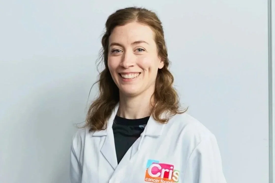
Dr Rebecca Ling
-
Researcher: Dr Casmir Turnquist
Institution: University of Oxford, Oxford
Cancers affecting children, adolescents, and young adults are not the same as those seen in adults. Although they are less common, they are often more difficult to diagnose, have fewer treatment options, and, in many cases, are considered rare cancers. This makes clinical decision-making more complex and prevents many patients from benefiting from targeted therapies.
One of the reasons is that these tumors are driven by very specific alterations in DNA, which act like faulty switches within cells and cause cancer to grow uncontrollably. Detecting these alterations quickly and accurately is key to choosing the right treatment, but today not all hospitals have the necessary tools, and results can take too long.
Improving diagnosis not only helps treat cancer more effectively: it also avoids unnecessary treatments, reduces toxicity, and allows doctors to act earlier, when therapeutic options are greater.
The Project
Cancers affecting children, adolescents and young adults are often rare, difficult to diagnose, and associated with fewer therapeutic options, largely because they are driven by highly specific genetic alterations that are not always detected in time. This project, led by Dr Casmir Turnquist, aims to transform the diagnosis of these tumours through the development of rapid and accurate tools capable of identifying key alterations directly in tumour samples.
By combining advanced genetic analyses, single-cell–level investigation and integration within clinical workflows, the research seeks to accelerate access to personalised treatments, reduce unnecessary toxicity, and improve survival and quality of life for children and young people with cancer.
Dr Casmir Turnquist
-
Researcher: Dr Eleni Louka
Institution: University of Oxford, Oxford
Project description: Juvenile myelomonocytic leukaemia (JMML) is a childhood leukaemia with a very poor prognosis, with limited therapeutic options beyond bone marrow transplantation and a high rate of relapse. This project, led by Dr Louka, focuses on studying a specific subpopulation of tumour cells identified as being responsible for these relapses, with the aim of developing more targeted, effective and less aggressive therapies that can improve treatment outcomes and prognosis for children with JMML.
Dr Eleni Louka
UCL / Great Ormond Street Institute
CRIS Cancer Foundation supports pioneering paediatric cancer research by funding innovative therapies and clinical trials at the UCL Great Ormond Street Institute of Child Health and Great Ormond Street Hospital. Through this partnership, CRIS Cancer accelerates the development of advanced immunotherapies, gene-editing techniques, and precision treatments for children with the most difficult-to-treat cancers.
-
Researcher: Persis Amrolia, Professor and Head of Bone Marrow Transplant
Centre: GOSH, London
Introduction
CAR-T therapies have revolutionised cancer treatment, especially for tumours that originate in blood cells such as leukaemias and lymphomas. They involve genetically engineering lymphocytes (the immune system cells responsible for destroying tumours) with a kind of radar or detector that helps them find and destroy cancer cells.
One of the most common leukaemias, and one that has benefited greatly from the emergence of CAR-T cells, is acute lymphoblastic leukaemia, especially in children. However, although more than 85% of children improve with the therapy, or even achieve complete remission, relapses are very common, and only 50% of these patients survive more than one year after treatment.
There are two main reasons for this. First, the molecular detectors introduced into the lymphocytes identify only a single molecule on the surface of tumour cells (in the case of acute lymphoblastic leukaemia, this is called CD19). If, over time, the tumour cells hide this molecule from their surface, the lymphocytes can no longer detect them, and the therapy stops working.
Secondly, these molecular detectors send a powerful signal to the lymphocytes when they encounter a tumour cell, causing the lymphocyte to attack and destroy it. However, it has been shown that such a strong signal can overstimulate the lymphocytes, causing them to become exhausted quickly and, once again, the therapy stops working.
Unless these two problems are solved, survival rates for children treated with CAR-T will not improve. For this reason, the clinical trial led by Dr Persis Amrolia aims to tackle both issues with current CAR-T therapies in order to prevent relapses.
The Project
In this clinical trial, Dr Persis Amrolia’s team will treat 12 patients using special CAR-T cells that have been modified in two ways:
They do not only recognise one tumour molecule (CD19), but also a second one (CD22). This means that if tumours hide one of them, the therapy may still continue to work.
They do not send such a strong signal to the lymphocytes when they encounter tumour cells. This means the lymphocytes are not overstimulated, do not become exhausted as quickly, and can continue working for longer.
With this trial, major progress is being made in refining therapies that are changing the way cancer is understood and treated.
Recent Progress
Dr Amrolia’s team has been completing all the requirements necessary to begin the clinical trial and enrol patients. Any therapy that is to be used in humans for the first time must meet a range of prior requirements, submit extensive documentation, and be approved by the relevant regulatory agencies and ethics committees.
For example, among many other things, they had to demonstrate on three separate occasions that they were able to produce the therapy under high-quality conditions, ensuring that the treatments given to children are always created as accurately and precisely as possible. After exhaustive work, all the necessary approvals have now been secured, allowing the project to begin.
Thanks to this approval, Dr Amrolia’s team has been able to open the trial and begin treating the first patients. So far, two children with high-risk leukaemias who had not responded to several previous therapies have already joined the trial. Over the course of the year, the team hopes to continue helping patients with these aggressive leukaemias and to report the first results of the trial.
Prof Persis Amrolia
-
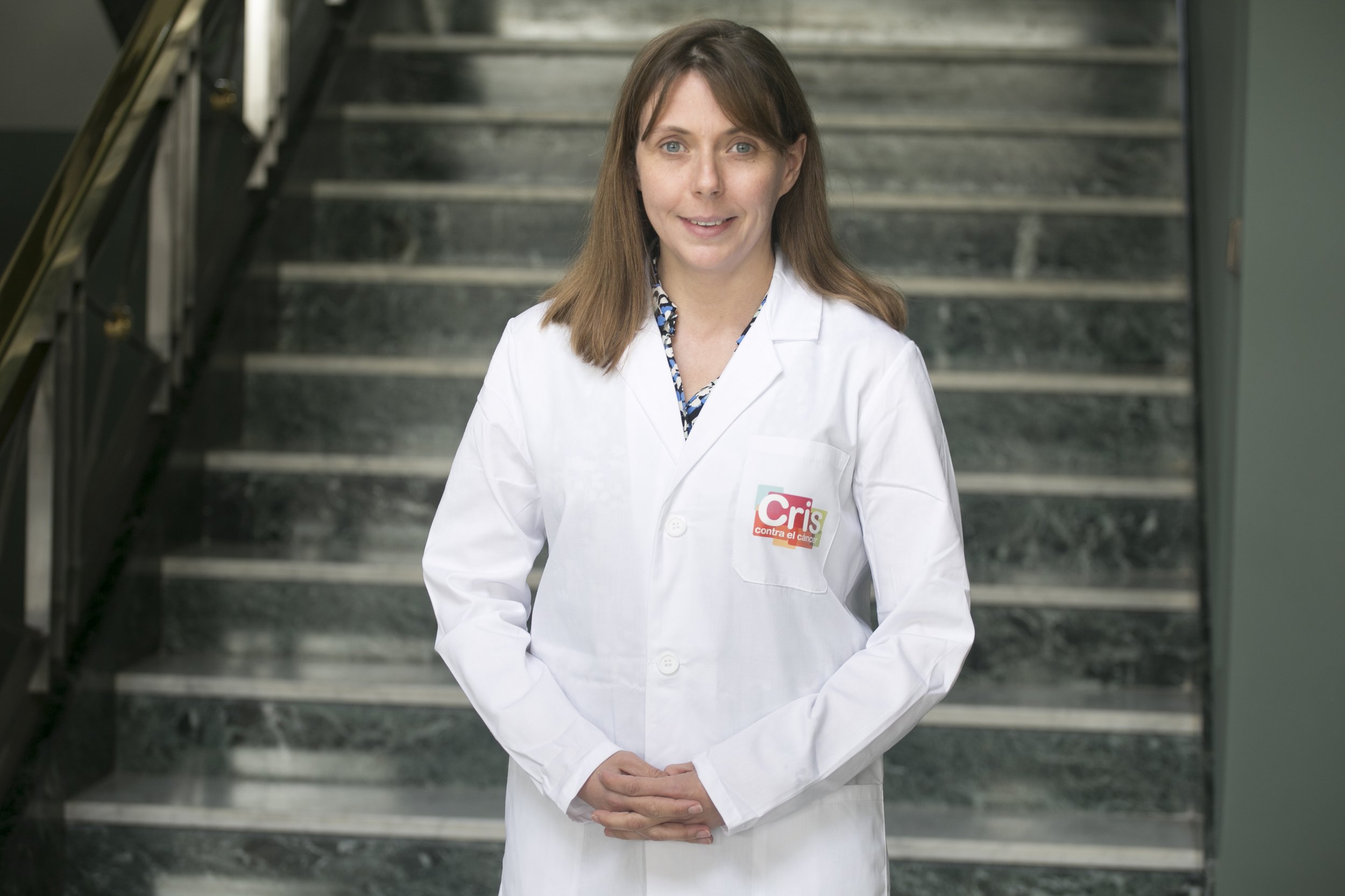
Researchers: Assoc Prof Karin Straathof, Consultant
Centre: UCL/GOSH, London
Introduction
Diffuse midline glioma (DMG) is a rare type of brain cancer that occurs more commonly in children than in adults. It develops in the central region of the brain, between the brainstem and surrounding structures, from which it takes its name. At present, these tumours are unfortunately incurable and carry a devastating prognosis. In fact, most children die within the first year following diagnosis, and fewer than 10% survive beyond two years, creating an urgent need for innovative therapies. One of the main challenges is that standard treatments, such as radiotherapy, only minimally extend patients’ lives. Although CAR-T immunotherapy has been effective in some childhood cancers, particularly blood cancers, it has not yet been proven safe or effective in brain tumours such as DMG.The Project
In this clinical trial, led by Dr Karin Straathof at the Great Ormond Street Institute of Child Health in London, CAR-T technology will be used for the first time to target these tumours. This approach involves collecting specific immune cells from the patient (T lymphocytes) and genetically engineering them to carry a molecular detector that enables them to identify and destroy tumour cells.In this case, the introduced detector allows the cells to target those carrying the GD2 molecule, which is commonly found on cells from this type of glioma but is rarely present on healthy cells.
Although it is still too early to evaluate the treatment’s effectiveness, the safety of the therapy has been demonstrated. In addition, researchers are studying whether the CAR-T cells successfully reach the tumour, how they behave once there, and what occurs at an immunological level when they attack tumour cells. This knowledge could help develop improved therapies in the future and support the design of larger clinical trials capable of benefiting more patients.
The trial is therefore progressing well. If the results meet expectations, this could represent the first step along a largely unexplored path: the development of safe and effective CAR-T therapies for a tumour type that currently has no effective treatments.
Thanks to the support of CRIS and GOSH CC, this pioneering project — the first clinical trial in Europe to use CAR-T therapy against a brain tumour — could open the door to far more effective treatments for a cancer that currently has no viable therapeutic options.

Assoc. Prof Karin Straathof